
Advantages
- Spheroids can be generated from established cell lines within two weeks.
- The spheroids exhibit brush border structures and express AQP1 and AQP3.
- Enables TGF-β-induced renal tubular fibrosis modeling, providing a useful platform for investigating disease mechanisms and drug efficacy evaluation.
Key Data
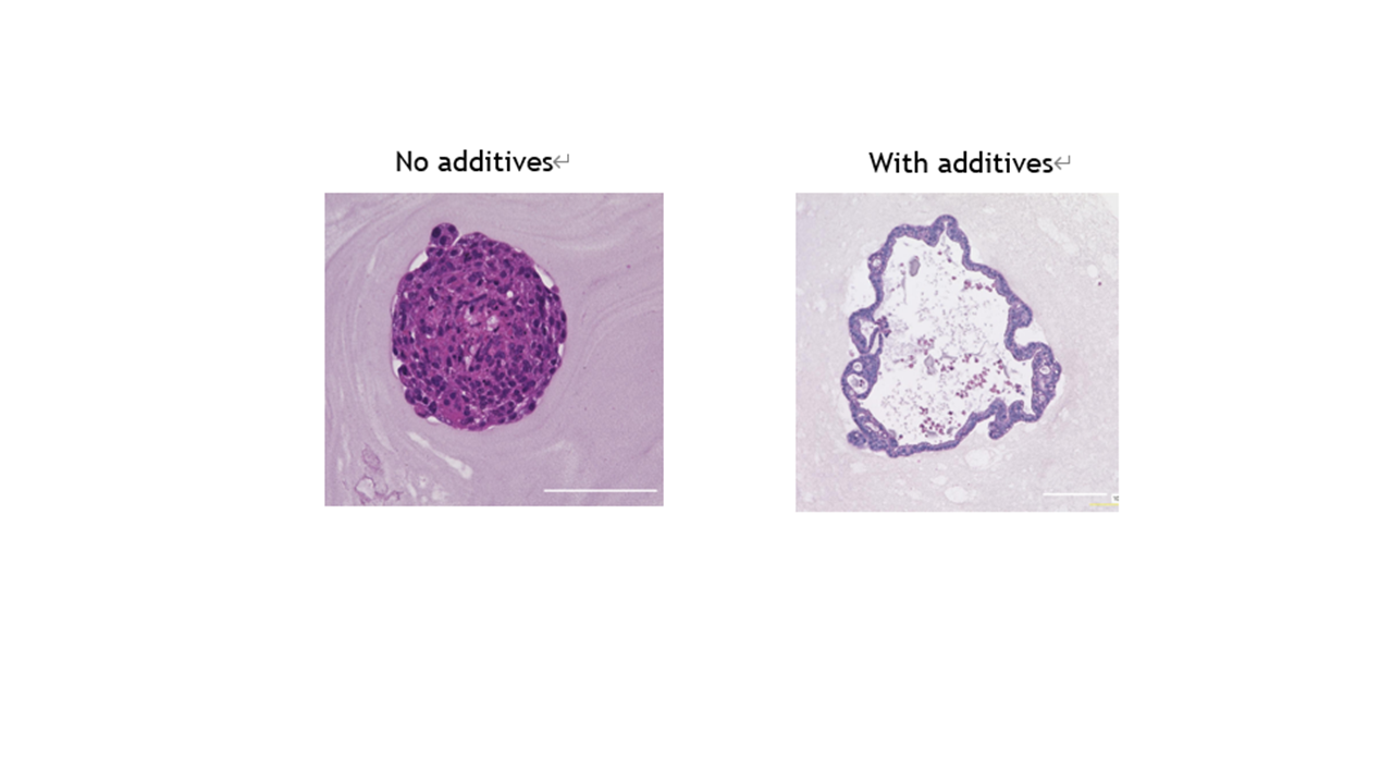
By adding the matrix main components of this process, a tubule structure was constructed.
On the left is the matrix without the main component added; on the right is the added CRFK. By addition, the structure of the tubule lumen is represented, and the expression of functional indicators (cadherin, aquaporin) as a tubule has been confirmed, making it possible for use in research and drug development.
Partnaring Model
This technology envisions diverse alliances and collaborations with animal drug manufacturers, CROs, and research tool manufacturers.
[For Veterinary Drug Manufacturers]
Joint research and technology provision as a screening platform for feline CKD treatments and renoprotective agents, as well as for toxicity and efficacy evaluation.
[For CRO]
Technology transfer and partnership to introduce the "Tubule Fibrosis Model" and "Urolithiasis and Polycystic Kidney Disease Model" as contract testing services using TGF-β stimulation into the contract portfolio.
[For Research Tool Manufacturers]
Joint development and commercialization of extracellular matrix-based culture materials required for 3D culture, as well as dedicated kits and assay plates for kidney spheroid production.
While contributing to the 3R principles as a biological replacement model, we can also contribute to licensing, joint research, and kit development to support new businesses and product development for the growing pet medical market. We would be happy to start with a detailed technical explanation and discussion.
Background and Technology
Chronic kidney disease (CKD) in cats is a major cause of morbidity and mortality, and the development of treatments is urgently needed. However, conventional feline renal cell lines (such as CRFK) have undergone mesenchymal transformations, and there has been no in vitro model reflecting the structure and function of the renal tubules of the living feline kidney. Furthermore, from the perspective of animal research ethics, reducing and replacing animal experimentation is strongly demanded by society.
This time, by three-dimensional culture of established feline renal cell lines such as CRFK cells in media supplemented with extracellular matrix (ECM) components (such as Matrigels containing laminin and collagen IV), we succeeded in producing kidney spheroids with a "tubule-like (lumen-containing) structure" in cats, which has been challenging to achieve, making it feasible as models and evaluation systems. This spheroid features brush border structures found in proximal tubules in vivo and clearly expresses aquaporins (AQP1 and AQP3) and the epithelial marker E-cadherin (CDH1). Furthermore, since it shows minimal spontaneous fibrotic activation, it enables clear evaluation with minimal background noise. This technology enables the provision of a functional feline kidney evaluation model through a relatively simple and rapid procedure.
Principal Investigator
Kotaro Nishi, Assistant Professor
Department of Veterinary Medicine, Faculty of Veterinary Medicine, Azabu University
Patents
Pending (domestic phase)