Advantages
(1) Captures the true extent of the tumor with higher precision than prior art (which used only contrast-enhanced T1 images) by inputting three types of MRI information into the AI: T1-weighted, T2-weighted, and contrast difference images.
(2) Obtains tumor activity information from standard MRI scans, enabling precise diagnosis at facilities without PET equipment.
(3) The signal values of the generated images strongly correlate with the tumor cell density in pathological tissue, directly contributing to improved accuracy in surgical and radiation therapy planning.
Key Data
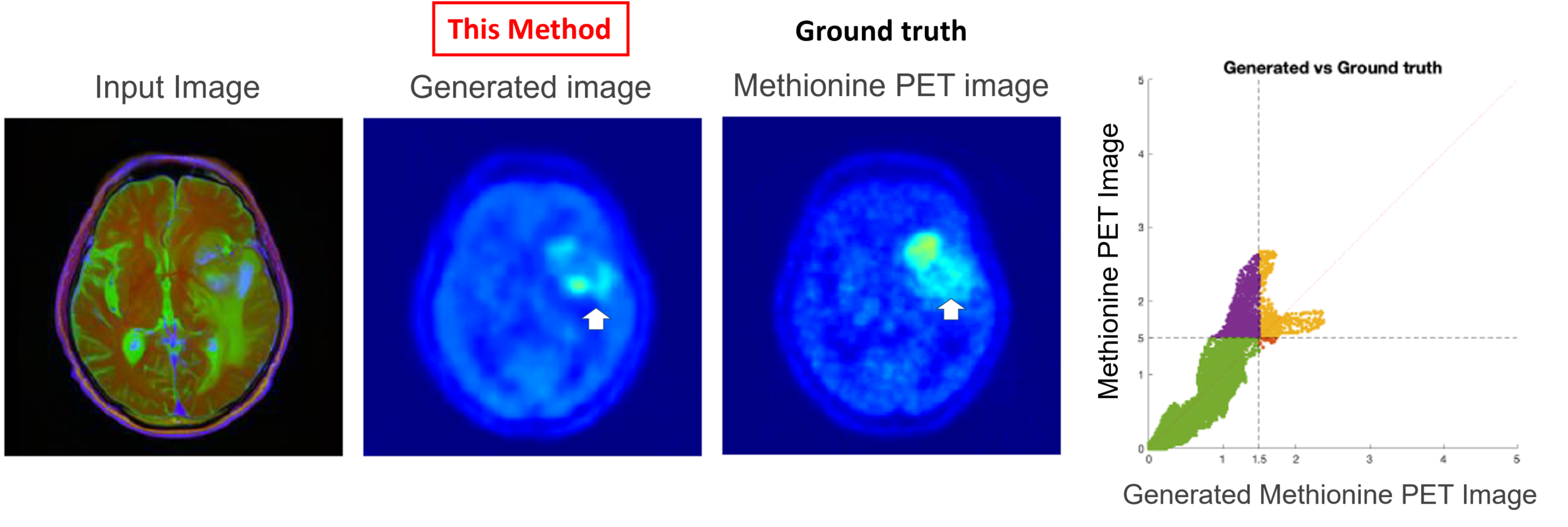
After homogenizing and performing difference processing on T1-weighted, contrast-enhanced T1-weighted, and T2-weighted images, generated methionine PET images were obtained using the Pix2Pix algorithm, resulting in highly accurate virtual PET images.
Partnaring Model
We believe this technology can be integrated as software into existing medical systems.
Through licensing agreements or joint research, we hope to collaborate with companies developing PACS viewers, surgical navigation systems, radiation therapy planning software, and surgical simulation software.
Disclosure of unpublished data is possible upon concluding a non-disclosure agreement with Asahikawa Medical University, and direct meetings with the researchers can also be arranged.
Background and Technology
The Problem: In glioblastoma treatment, "invisible cancer" cells infiltrate tissue beyond what standard MRI can detect, leading to tumor recurrence after surgery.
A Flawed Solution: Methionine PET scans can visualize this invisible infiltration, but the technology is too expensive and scarcely available for routine use.
The New Technology: An AI-powered technology has been developed to generate high-precision, virtual methionine PET images using only standard MRI scans.
How It Works: The AI achieves high accuracy by using a multi-faceted input of three MRI data types (T1-weighted, T2-weighted, and contrast difference images) combined with a proprietary preprocessing technique.
The Benefit: This enables non-invasive, low-cost, and precise tumor diagnosis at any facility with an MRI, making advanced treatment planning accessible and improving patient outcomes.
Principal Investigator
Dr Manabu Kinoshita (Asahikawa Medical University School of Medicine)
Patents and Publications
Patent pending (unpublished)