
Advantage and Core Benefit
●High Accuracy, Zero Damage:
96–99%+ accuracy via CNN on a single 2D image of live cells (Hoechst) or tissue sections (DAPI). Adherent cells remain intact without detachment.
●Cost & Time Efficiency:
Eliminates expensive workflows (specific antibodies, FACS, scRNA-seq). Uses only cheap, standard DNA fluorescent dyes.
●Broad Applicability:
Proven on neurons, fibroblasts, neural stem cells, chondrocytes, and lens cells.
Background and Technology
Conventional cell identification (immunostaining, FACS) relies on costly markers and risks altering adherent cells through detachment.
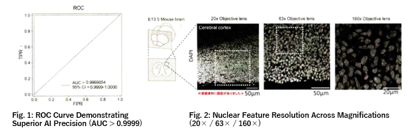
This breakthrough technology leverages macro-DNA arrangement (heterochromatin variations) within the nucleus as a cell-type-specific signature. Applying a CNN to 2D grayscale nuclear images (single-plane, confocal microscopy, ~160x objective) enables highly accurate, marker-free classification.
Data (Mouse-derived)
Achieves precision beyond human visual limits:
●Live Cells (Hoechst): >99% average accuracy
・Tested on 4 cell types (e.g., Neurons: 99.88%, P19: 100%).
・Identified unlearned ES-derived neurons at 99.25%.
●Fixed Tissue (DAPI): >96% average accuracy
・Tested on 4 cranial cell types (e.g., Neural Stem Cells: 97.95%, Neurons: 96.95%).
Partnership Expectations
Seeking commercialization partners in:
●Regenerative Medicine (Cell Sheets, etc.): Non-destructive, inline QA/QC for cell uniformity and contamination.
●Pharma & CROs: Antibody-free, rapid cell profiling of pathological tissue sections (e.g., tumor microenvironments).
●Microscopy & Optics: Bundled AI software (automated segmentation/classification) for microscopes.
Collaboration Model:
Technical/Academic Advisory. Partners lead development, while the researcher provides proprietary know-how (e.g., optimizing image parameters, adapting to lower magnifications). Guidance for expanding from mouse data to human cells (e.g., iPS-derived neurons) is fully supported.
Principal Investigator
Prof. Tetsuichiro Saito(Graduate School of Medicine, Chiba University )
Patents and Publications
WO2026160417
Information on related literature is included in the materials. Please click the “Iinterested in this technology? [Download Materials]” button below to download and view the materials.